Not everything in the brain is meant to last. As our brains assemble, trillions of neural connections have to be built or torn down at the right time and place. Otherwise, the seeds of disorders like autism can take root. Cold Spring Harbor Laboratory Assistant Professor Gabrielle Pouchelon studies how the brain is wired early in life. In doing so, she hopes to find the origins of various brain dysfunctions and new ways to treat them.
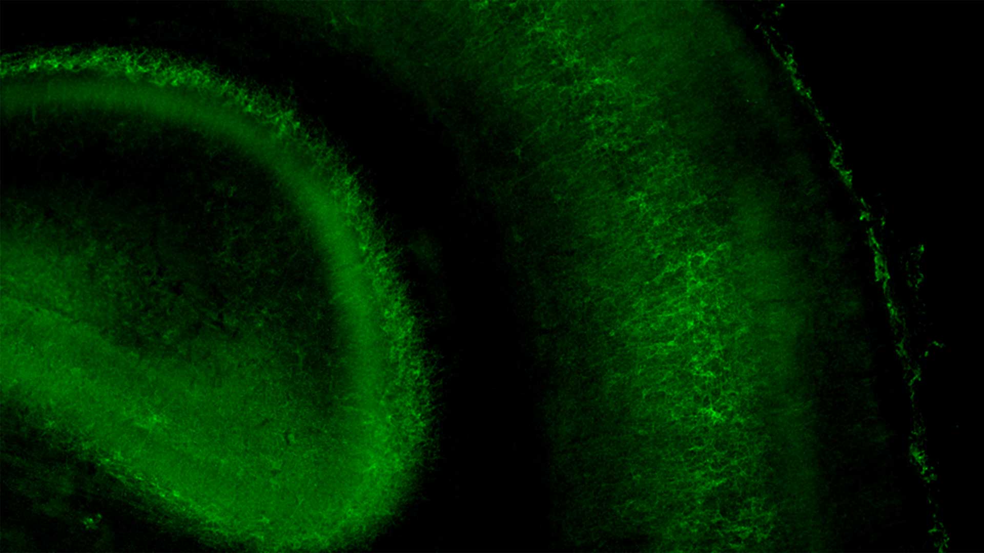
In a new study, Pouchelon and her team zero in on a process known as pruning. This is when the brain removes unnecessary connections between neurons. The pruning of long-lasting connections is relatively well-known. Pouchelon’s team focuses on special early connections that get cut to make way for long-lasting circuits in the mature brain. Though temporary, these early connections may play a critical role in shaping developing brain circuits.
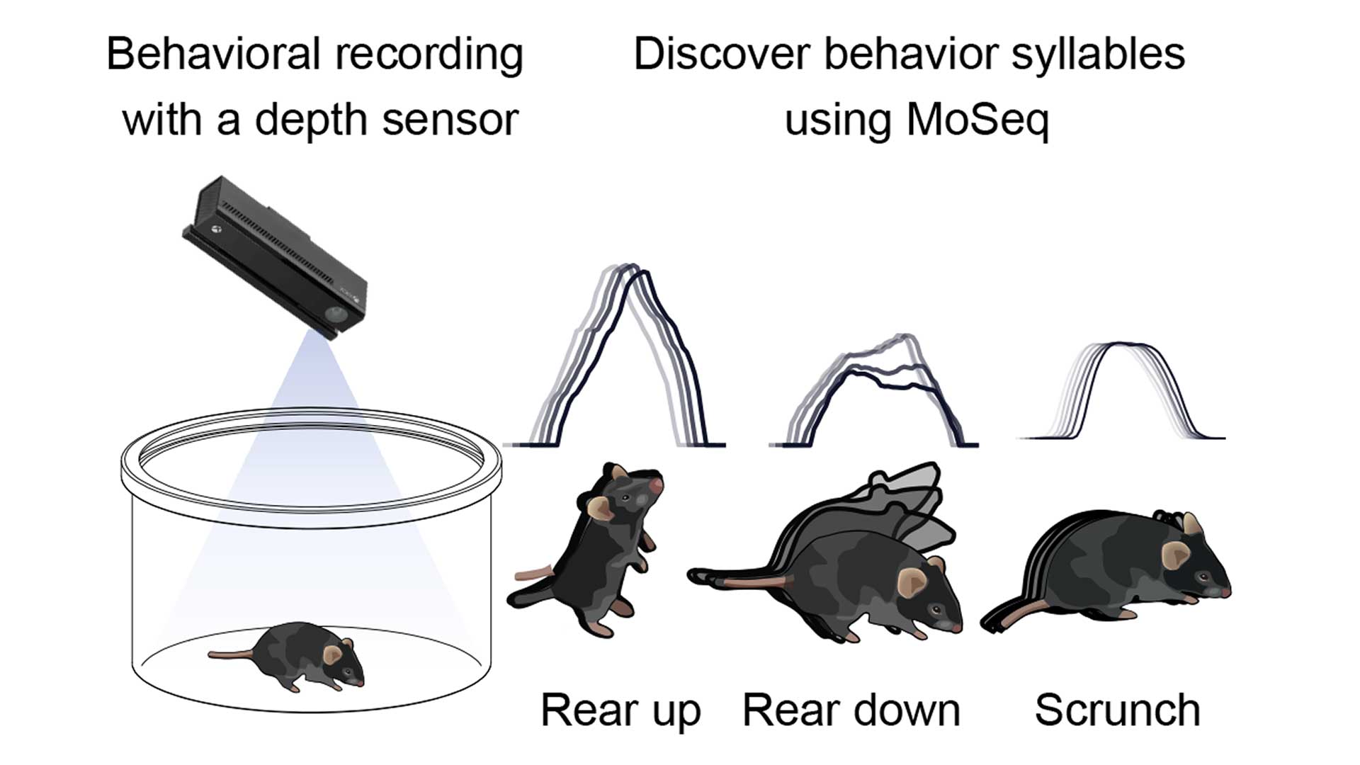
Pouchelon’s lab has now discovered that a receptor protein named mGluR1 helps regulate the timing of these temporary connections in the mouse brain. Her team found that without mGluR1, neural connections stick around too long in the brain region that controls and processes touch via the whiskers. When the sensory circuit fails to mature properly, the mice demonstrate atypical behaviors. For example, they don’t stand on their hind legs and sniff around the way other mice do.
Importantly, the team notes that this critical step in circuit development occurs during the first week after birth. “The way the receptor works seems to be different than what has been described in adulthood,” Pouchelon says. She explains:
“In the context of neurodevelopmental disorders, that means when we try to target developmental defects, we could have a totally different therapeutic effect at different stages during development.”
Pouchelon’s team hopes their discovery may serve as a guide for designing future therapeutics to treat brain dysfunction early. “The brain is a wonderful machine whose job is to adapt,” says Dimitri Dumontier, the postdoc in Pouchelon’s lab who co-led this study. “So, when you study neurodevelopmental disorders in adults or even teenagers, it is difficult to identify which mechanisms are causing the symptoms. That is why understanding early milestones of brain development is key.”
The hope is that by figuring out exactly how the brain matures, scientists can rescue this process early. This could help prevent symptoms of neurological disorders like autism from showing up in the first place. After all, the world is difficult enough to navigate as is. Pouchelon and Dumontier’s work could someday help make life easier for countless young people.
Written by: Luis Sandoval, Communications Specialist | sandova@cshl.edu | 516-367-6826
Funding
European Molecular Biology Organization, Swiss National Science Foundation, Hearst Foundations, The Pershing Square Foundation, National Institutes of Health, Simons Foundation
Citation
Dwivedi, D., Dumontier, D., et al., “Metabotropic signaling within somatostatin interneurons controls transient thalamocortical inputs during development”, Nature Communications, July 26, 2024. DOI: 10.1038/s41467-024-49732-w
Core Facilites
Principal Investigator
Gabrielle Pouchelon
Assistant Professor
Ph.D., University of Geneva – Switzerland, 2013